Thu, Oct 15, 2026
Volume 12 - Contnious Publishing
Iran J Neurosurg 2026, 12 - Contnious Publishing: 0-0 |
Back to browse issues page



Download citation:
BibTeX | RIS | EndNote | Medlars | ProCite | Reference Manager | RefWorks
Send citation to:


BibTeX | RIS | EndNote | Medlars | ProCite | Reference Manager | RefWorks
Send citation to:
Parajiya R, Patil A, Deshmukh P, Goudar P. Acute Subdural Hematoma Without Trauma: An Uncommon Presentation of Middle Cerebral Artery Aneurysm: Case Report. Iran J Neurosurg 2026; 12 : 6
URL: http://irjns.org/article-1-509-en.html
URL: http://irjns.org/article-1-509-en.html


1- Department of Neurosurgery, Jawaharlal Nehru Medical College, Belagavi, India.
2- Department of Anesthesiology, Jawaharlal Nehru Medical College, Belagavi, India ,drabhishekpatil87@gmail.com
3- Department of Anesthesiology, Jawaharlal Nehru Medical College, Belagavi, India
4- Department of Radiology, Jawaharlal Nehru Medical College, Belagavi, India.
2- Department of Anesthesiology, Jawaharlal Nehru Medical College, Belagavi, India ,
3- Department of Anesthesiology, Jawaharlal Nehru Medical College, Belagavi, India
4- Department of Radiology, Jawaharlal Nehru Medical College, Belagavi, India.
Keywords: Middle cerebral artery aneurysm, spontaneous subdural hematoma, non-traumatic subdural hemorrhage, Vascular imaging in SDH, CT angiography in SDH
Full Text [PDF 1039 kb]
(176 Downloads)
| Abstract (HTML) (745 Views)
Full Text: (72 Views)
1. Background and Importance
Acute subdural hematoma (ASDH) is classically traumatic, resulting from torn bridging veins. By contrast, spontaneous ASDH represents 0.7–6.7% of all ASDH cases, with many series reporting approximately 2.5%. Within this niche category, ruptured saccular aneurysms account for 0.5–7.9% of all aneurysmal bleeds. Middle cerebral artery (MCA) aneurysms contribute roughly 24–27% of aneurysmal ASDH cases, second only to PcomA lesions (approximately 30%). Because routine trauma protocols often overlook vascular imaging, aneurysm-related ASDH is easily misdiagnosed, delaying surgical clipping or coiling and worsening outcomes [1-5].
Current concerns include the lack of clear imaging algorithms for atraumatic ASDH, the potential for misdiagnosis as venous bleeding, and the limited awareness of aneurysms as a cause of subdural hematoma. Early recognition is paramount. Patients deteriorate quickly due to mass effect, rebleeding, or occult vasospasm. Surgical evacuation alone, without aneurysm occlusion, risks catastrophic re-rupture. The dual goals are therefore: (1) decompressing the hematoma to reverse herniation and (2) permanently securing the aneurysm [6, 7]. This case underscores the importance of early vascular imaging and contributes to ongoing efforts to refine diagnostic and management strategies for this uncommon but critical condition.
2. Case Presentation
A 48-year-old female with no history of trauma, coagulopathy, or hypertension presented to the emergency department with sudden-onset headache for 2 days, giddiness, and right-sided hemiparesis. There was no history of recent falls, seizures, or anticoagulant use.
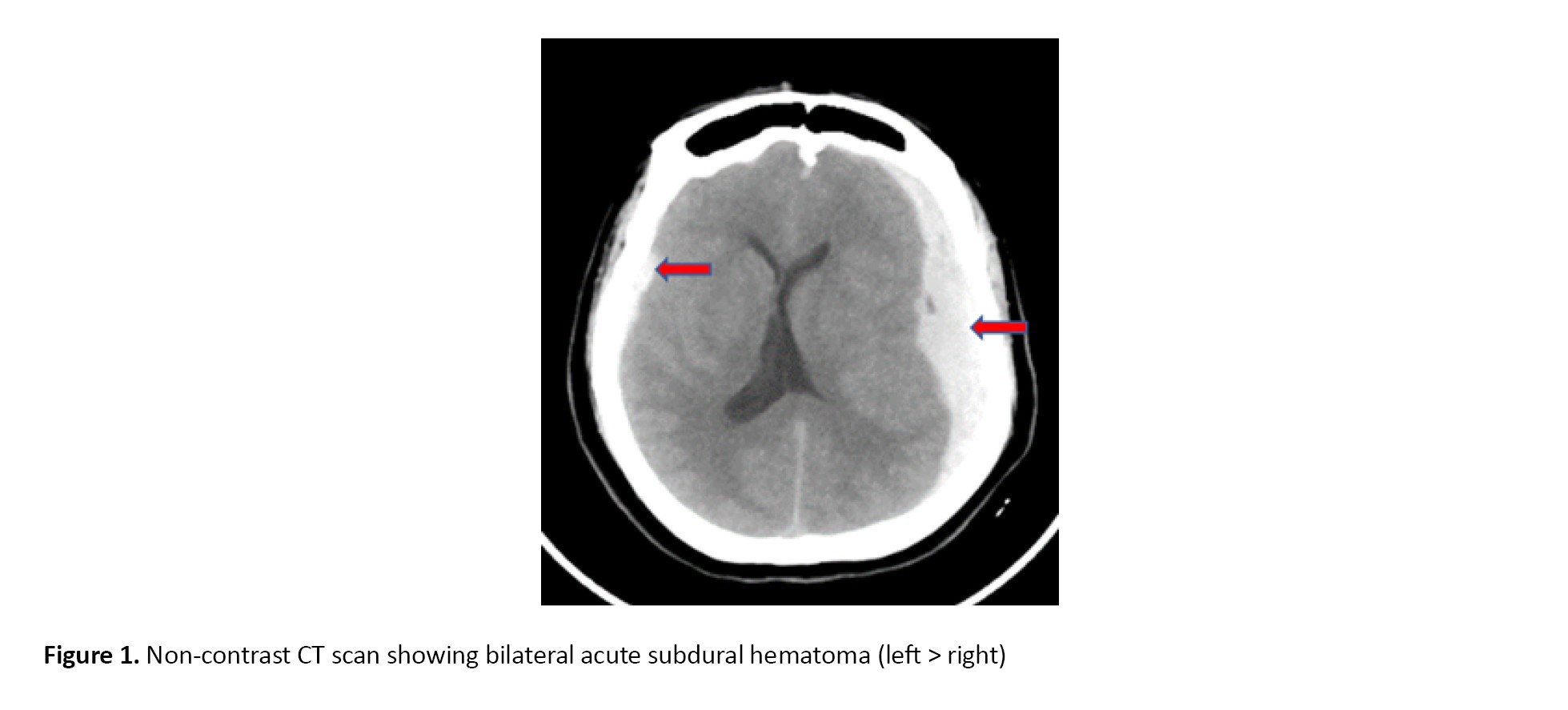
Neurological examination: Glasgow coma scale (GCS)score was 14/15 with equal pupil reactions and right-sided hemiparesis 3/5 (Medical Research Council scale). Non-contrast computed tomography (CT) brain revealed bilateral ASDH (left > right) with midline shift to the right without obvious subarachnoid hemorrhage (Figure 1).
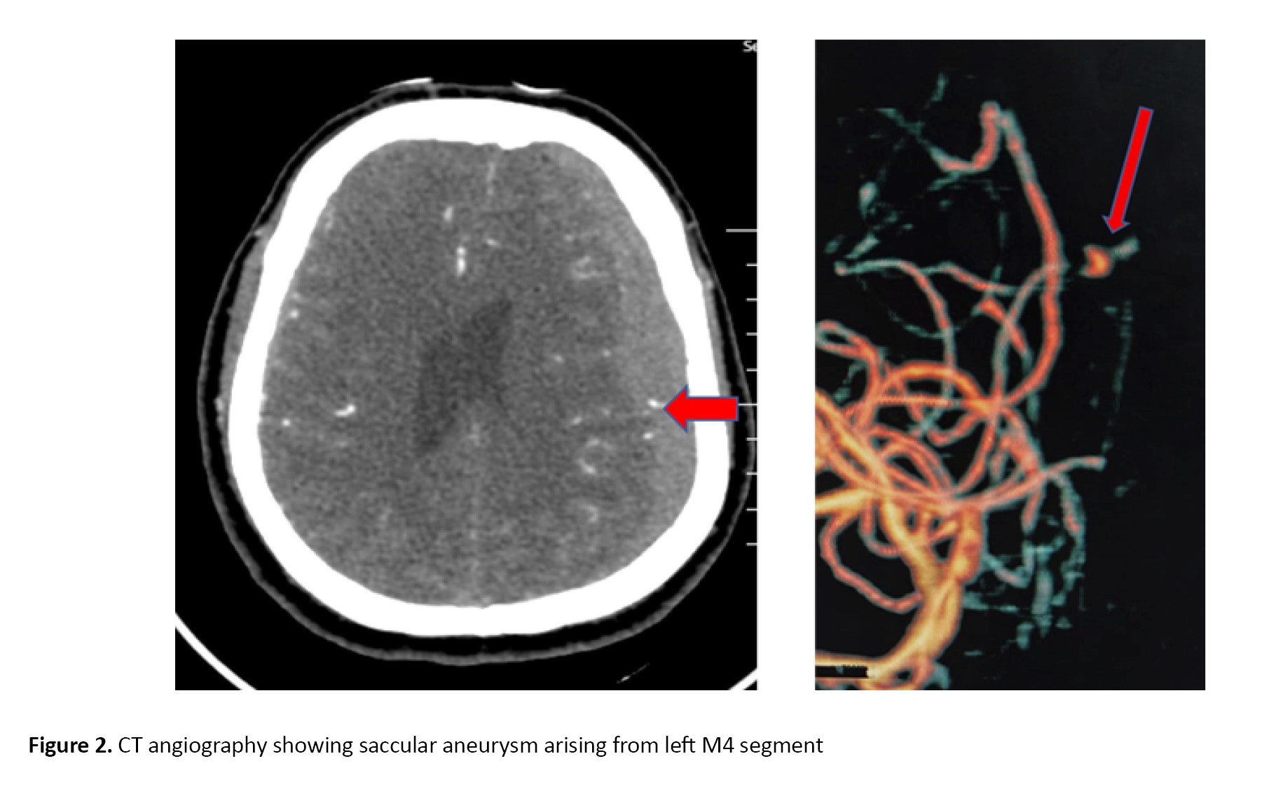
 CT angiography revealed a saccular aneurysm arising from the left M4 segment with active extravasation of contrast material, with the aneurysm dome embedded within the subdural hematoma (Figure 2).
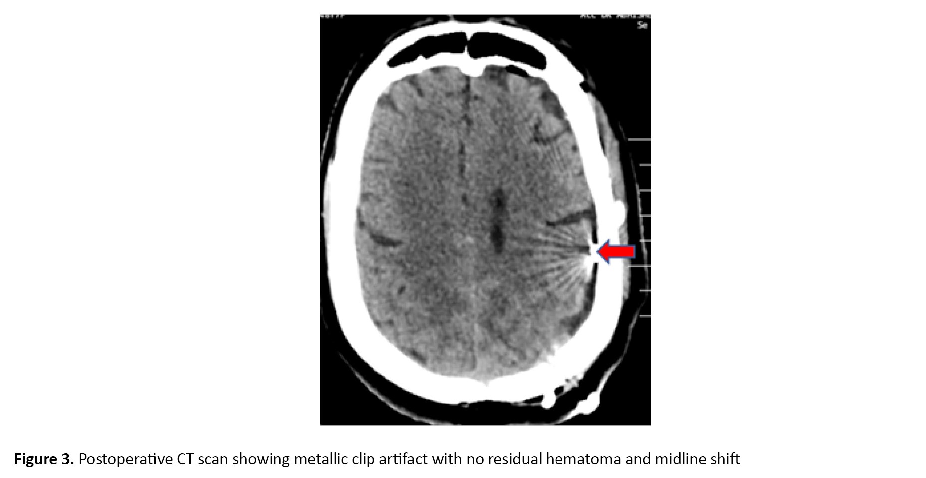
 The patient underwent left fronto-temporo-parietal craniotomy with hematoma evacuation and aneurysm clipping. Intraoperatively, active bleeding from an aneurysm in the M4 segment was identified. The ruptured aneurysm was successfully clipped under microscopic visualization with a 3 mm straight clip. Postoperative scan showed complete resolution of previously observed hyperdense ASDH over the bilateral cerebral convexity and reduction in midline shift, and no ischemic change or rebleed was noted. Metallic clip artifact was observed (Figure 3).
 In the present case, no evidence of infection or systemic illness was found; blood cultures and echocardiography were negative, and the aneurysm morphology was saccular rather than fusiform, arguing against a mycotic or traumatic pseudoaneurysm. The absence of prior head trauma further supports an idiopathic saccular origin.
Postoperatively, neurological status improved steadily, with resolution of hemiparesis and normalization of sensorium. By postoperative day 4, the patient was able to mobilize independently. At 3-month follow-up, she returned to daily activities with a modified Rankin scale (mRS) score of 1, indicating minimal symptoms and excellent functional recovery.
Timing of computed tomography angiography (CTA) and surgical intervention
Given the patient’s neurological deterioration and radiological evidence of significant mass effect, the decision to perform CTA was made immediately upon presentation. CTA was completed within one hour of hospital arrival, and the on-call radiologist promptly confirmed the diagnosis of a ruptured distal M4 aneurysm. The patient was then taken directly to the operating room without further delay. This brief imaging interval did not prolong the time to surgical decompression but enabled definitive aneurysm identification and intraoperative planning.
3. Discussion
Spontaneous aneurysmal ASDH shows a marked female predominance, with ~70% of cases occurring in patients with a mean age of approximately 48 years, though reported cases span a wide age range from 15 to 85 years [1, 8]. Regarding aneurysm location, the posterior communicating artery (PcomA) is most frequently implicated, accounting for 30–39% of cases. MCA follows this at 24–27%, anterior communicating artery (AcomA) at 13–15%, and distal anterior cerebral artery at 9–11%, with other locations comprising the remaining 12%. This distribution has remained stable over decades [1, 3]. Hematomas most often involve the cerebral convexity (58%), followed by interhemispheric (22%) and tentorial (17%) sites; rare posterior fossa or spinal cases likely result from caudal blood descent [3, 9, 10].
Why distal MCA aneurysms favor subdural spread
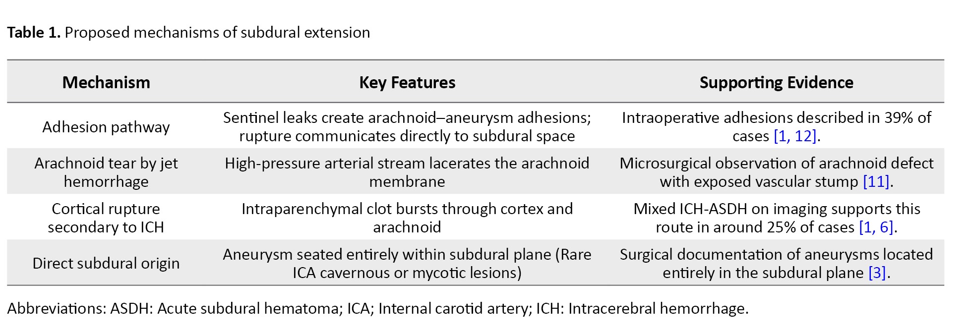
The distal MCA is close to the subdural space, making aneurysms prone to rupture through the arachnoid and bleeding directly into it. Lateral or superior dome projection increases this risk. Prior minor bleeds may cause arachnoid–dural adhesions, creating a low-resistance path for arterial blood, leading to spontaneous ASDH without trauma [1, 11]. Table 1 presents mechanisms involved in subdural hematoma secondary to aneurysmal etiology.
The proposed mechanism in our case involves localized arterial wall weakness at branching points, possibly aggravated by hemodynamic stress in distal cortical vessels.
Clinical presentation
Patients with aneurysm-related spontaneous ASDH frequently report sentinel headaches, with retrospective studies showing that 15-40% of aneurysmal subarachnoid hemorrhage cases recall such warning headaches in the days to weeks preceding rupture. These headaches may reflect minor adhesion-forming bleeds prior to catastrophic hemorrhage [1, 12]. At presentation, approximately 50% of patients are in Hunt and Hess grade IV–V, often manifesting anisocoria and hemiparesis, warning signs of impending herniation; poor-grade presentations are associated with high morbidity and mortality [7]. Most patients had no history of trauma, supporting a non-traumatic origin in the majority of these cases, although specific series quantifying this rate are limited [13].
Radiology
On non-contrast CT, the hematoma typically appears as a hyperdense crescentic collection over one cerebral hemisphere, often crossing sutures. Additionally, a disproportionate midline shift relative to the hematoma’s thickness should raise suspicion for an arterial source rather than venous injury [14].
CTA sensitivity varies significantly across recent studies, ranging from 70% to 95% overall, with markedly reduced sensitivity for smaller aneurysms (<3-5 mm), and allows rapid 3D reconstruction when time constraints limit digital subtraction angiography (DSA) [2, 12]. However, CTA is less reliable in identifying thrombosed or very distal cortical aneurysms, and repeat imaging within 24 hours is often recommended if initial CTA is negative and suspicion remains high [14, 16].
DSA is superior, particularly valuable for detecting small, thrombosed, or distal aneurysms that may be missed on CTA [17]. In equivocal cases, magnetic resonance angiography with fluid-attenuated inversion recovery or susceptibility-weighted imaging can detect subtle subarachnoid blood or “pseudo-subarachnoid hemorrhage (SAH)” signs, helping identify aneurysmal sources [16].
Management and prognostication
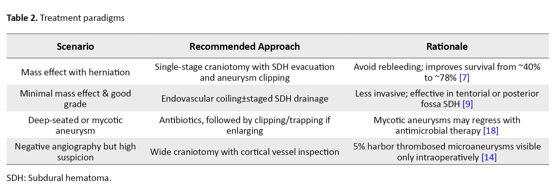
Table 2 describes recommended treatment protocols for ASDH across different presentations.
Early series of aneurysm-induced spontaneous ASDH reported high mortality rates of around 50%, reflecting limited recognition and rudimentary treatment protocols at the time. In contrast, contemporary case series utilizing modern multimodal management, including early vascular imaging, timely surgical clipping or endovascular coiling, and neurocritical care, report much lower mortality rates of approximately 16–17% in aneurysmal subarachnoid hemorrhage cohorts, which likely subsume ASDH cases as well [4, 7].
Functional recovery also differs by hemorrhage pattern. Patients presenting with isolated ASDH (without subarachnoid hemorrhage) tend to have significantly better outcomes, with approximately 38% achieving favorable functional status (mRS 0–2); whereas those with mixed SAH and SDH presentations fare worse, with only around 6% reaching similar recovery levels. While direct series specific to ASDH are sparse, this pattern aligns with broader SAH outcome data [4, 19].
Predictors of poor outcomes include delayed diagnosis, especially when vascular imaging is deferred; Hunt and Hess grades IV–V at presentation; clinical signs of brainstem herniation (e.g. anisocoria, hemiparesis); and involvement of posterior circulation aneurysms, which have higher risks due to deeper location and often poorer grade at rupture [6, 20].
4. Conclusion
Spontaneous ASDH is a neurosurgical emergency that requires vigilance for an underlying ruptured aneurysm, especially along the cortical branches of the MCA. Clinical clues (atraumatic onset, severe headache, rapid decline in neurological status), imaging patterns (large convexity clot with minimal SAH), and immediate angiography converge to reveal the culprit. Optimal management includes early hematoma evacuation with definitive aneurysm occlusion, yielding survival exceeding 80% in modern series. Our report contributes to the expanding literature on MCA aneurysm–related ASDH, highlighting the crucial maxim: “Every unexplained ASDH warrants a vascular search.”
Ethical Considerations
Compliance with ethical guidelines
The patient provided written informed consent for the publication of this case report and any accompanying images.
Funding
This research did not receive any grants from funding agencies in the public, commercial, or non-profit sectors.
Authors' contributions
All authors contributed equally to the conception and design of the study, data collection and analysis, interpretation of the results, and manuscript drafting. Each author approved the submission of the final version of the manuscript.
Conflict of interest
The authors declared no conflict of interest.
References
Acute subdural hematoma (ASDH) is classically traumatic, resulting from torn bridging veins. By contrast, spontaneous ASDH represents 0.7–6.7% of all ASDH cases, with many series reporting approximately 2.5%. Within this niche category, ruptured saccular aneurysms account for 0.5–7.9% of all aneurysmal bleeds. Middle cerebral artery (MCA) aneurysms contribute roughly 24–27% of aneurysmal ASDH cases, second only to PcomA lesions (approximately 30%). Because routine trauma protocols often overlook vascular imaging, aneurysm-related ASDH is easily misdiagnosed, delaying surgical clipping or coiling and worsening outcomes [1-5].
Current concerns include the lack of clear imaging algorithms for atraumatic ASDH, the potential for misdiagnosis as venous bleeding, and the limited awareness of aneurysms as a cause of subdural hematoma. Early recognition is paramount. Patients deteriorate quickly due to mass effect, rebleeding, or occult vasospasm. Surgical evacuation alone, without aneurysm occlusion, risks catastrophic re-rupture. The dual goals are therefore: (1) decompressing the hematoma to reverse herniation and (2) permanently securing the aneurysm [6, 7]. This case underscores the importance of early vascular imaging and contributes to ongoing efforts to refine diagnostic and management strategies for this uncommon but critical condition.
2. Case Presentation
A 48-year-old female with no history of trauma, coagulopathy, or hypertension presented to the emergency department with sudden-onset headache for 2 days, giddiness, and right-sided hemiparesis. There was no history of recent falls, seizures, or anticoagulant use.
Neurological examination: Glasgow coma scale (GCS)score was 14/15 with equal pupil reactions and right-sided hemiparesis 3/5 (Medical Research Council scale). Non-contrast computed tomography (CT) brain revealed bilateral ASDH (left > right) with midline shift to the right without obvious subarachnoid hemorrhage (Figure 1).
Postoperatively, neurological status improved steadily, with resolution of hemiparesis and normalization of sensorium. By postoperative day 4, the patient was able to mobilize independently. At 3-month follow-up, she returned to daily activities with a modified Rankin scale (mRS) score of 1, indicating minimal symptoms and excellent functional recovery.
Timing of computed tomography angiography (CTA) and surgical intervention
Given the patient’s neurological deterioration and radiological evidence of significant mass effect, the decision to perform CTA was made immediately upon presentation. CTA was completed within one hour of hospital arrival, and the on-call radiologist promptly confirmed the diagnosis of a ruptured distal M4 aneurysm. The patient was then taken directly to the operating room without further delay. This brief imaging interval did not prolong the time to surgical decompression but enabled definitive aneurysm identification and intraoperative planning.
3. Discussion
Spontaneous aneurysmal ASDH shows a marked female predominance, with ~70% of cases occurring in patients with a mean age of approximately 48 years, though reported cases span a wide age range from 15 to 85 years [1, 8]. Regarding aneurysm location, the posterior communicating artery (PcomA) is most frequently implicated, accounting for 30–39% of cases. MCA follows this at 24–27%, anterior communicating artery (AcomA) at 13–15%, and distal anterior cerebral artery at 9–11%, with other locations comprising the remaining 12%. This distribution has remained stable over decades [1, 3]. Hematomas most often involve the cerebral convexity (58%), followed by interhemispheric (22%) and tentorial (17%) sites; rare posterior fossa or spinal cases likely result from caudal blood descent [3, 9, 10].
Why distal MCA aneurysms favor subdural spread
The distal MCA is close to the subdural space, making aneurysms prone to rupture through the arachnoid and bleeding directly into it. Lateral or superior dome projection increases this risk. Prior minor bleeds may cause arachnoid–dural adhesions, creating a low-resistance path for arterial blood, leading to spontaneous ASDH without trauma [1, 11]. Table 1 presents mechanisms involved in subdural hematoma secondary to aneurysmal etiology.
The proposed mechanism in our case involves localized arterial wall weakness at branching points, possibly aggravated by hemodynamic stress in distal cortical vessels.
Clinical presentation
Patients with aneurysm-related spontaneous ASDH frequently report sentinel headaches, with retrospective studies showing that 15-40% of aneurysmal subarachnoid hemorrhage cases recall such warning headaches in the days to weeks preceding rupture. These headaches may reflect minor adhesion-forming bleeds prior to catastrophic hemorrhage [1, 12]. At presentation, approximately 50% of patients are in Hunt and Hess grade IV–V, often manifesting anisocoria and hemiparesis, warning signs of impending herniation; poor-grade presentations are associated with high morbidity and mortality [7]. Most patients had no history of trauma, supporting a non-traumatic origin in the majority of these cases, although specific series quantifying this rate are limited [13].
Radiology
On non-contrast CT, the hematoma typically appears as a hyperdense crescentic collection over one cerebral hemisphere, often crossing sutures. Additionally, a disproportionate midline shift relative to the hematoma’s thickness should raise suspicion for an arterial source rather than venous injury [14].
CTA sensitivity varies significantly across recent studies, ranging from 70% to 95% overall, with markedly reduced sensitivity for smaller aneurysms (<3-5 mm), and allows rapid 3D reconstruction when time constraints limit digital subtraction angiography (DSA) [2, 12]. However, CTA is less reliable in identifying thrombosed or very distal cortical aneurysms, and repeat imaging within 24 hours is often recommended if initial CTA is negative and suspicion remains high [14, 16].
DSA is superior, particularly valuable for detecting small, thrombosed, or distal aneurysms that may be missed on CTA [17]. In equivocal cases, magnetic resonance angiography with fluid-attenuated inversion recovery or susceptibility-weighted imaging can detect subtle subarachnoid blood or “pseudo-subarachnoid hemorrhage (SAH)” signs, helping identify aneurysmal sources [16].
Management and prognostication
Table 2 describes recommended treatment protocols for ASDH across different presentations.
Early series of aneurysm-induced spontaneous ASDH reported high mortality rates of around 50%, reflecting limited recognition and rudimentary treatment protocols at the time. In contrast, contemporary case series utilizing modern multimodal management, including early vascular imaging, timely surgical clipping or endovascular coiling, and neurocritical care, report much lower mortality rates of approximately 16–17% in aneurysmal subarachnoid hemorrhage cohorts, which likely subsume ASDH cases as well [4, 7].
Functional recovery also differs by hemorrhage pattern. Patients presenting with isolated ASDH (without subarachnoid hemorrhage) tend to have significantly better outcomes, with approximately 38% achieving favorable functional status (mRS 0–2); whereas those with mixed SAH and SDH presentations fare worse, with only around 6% reaching similar recovery levels. While direct series specific to ASDH are sparse, this pattern aligns with broader SAH outcome data [4, 19].
Predictors of poor outcomes include delayed diagnosis, especially when vascular imaging is deferred; Hunt and Hess grades IV–V at presentation; clinical signs of brainstem herniation (e.g. anisocoria, hemiparesis); and involvement of posterior circulation aneurysms, which have higher risks due to deeper location and often poorer grade at rupture [6, 20].
4. Conclusion
Spontaneous ASDH is a neurosurgical emergency that requires vigilance for an underlying ruptured aneurysm, especially along the cortical branches of the MCA. Clinical clues (atraumatic onset, severe headache, rapid decline in neurological status), imaging patterns (large convexity clot with minimal SAH), and immediate angiography converge to reveal the culprit. Optimal management includes early hematoma evacuation with definitive aneurysm occlusion, yielding survival exceeding 80% in modern series. Our report contributes to the expanding literature on MCA aneurysm–related ASDH, highlighting the crucial maxim: “Every unexplained ASDH warrants a vascular search.”
Ethical Considerations
Compliance with ethical guidelines
The patient provided written informed consent for the publication of this case report and any accompanying images.
Funding
This research did not receive any grants from funding agencies in the public, commercial, or non-profit sectors.
Authors' contributions
All authors contributed equally to the conception and design of the study, data collection and analysis, interpretation of the results, and manuscript drafting. Each author approved the submission of the final version of the manuscript.
Conflict of interest
The authors declared no conflict of interest.
References
- Beucler N, Haikal C, Hibbert D, Sellier A, Joubert C, Desse N, et al. Spontaneous acute subdural hematoma: beware of the aneurysm. Journal of Neurosciences in Rural Practice. 2019; 10(3):537-41. [DOI:10.1055/s-0039-1697770] [PMID] [PMCID]
- Marbacher S, Tomasi O, Fandino J. Management of patients presenting with acute subdural hematoma due to ruptured intracranial aneurysm. International Journal of Vascular Medicine. 2012; 2012:753596. [DOI:10.1155/2012/753596] [PMID] [PMCID]
- Gong J, Sun H, Shi XY, Liu WX, Shen Z. Pure subdural haematoma caused by rupture of middle cerebral artery aneurysm: Case report and literature review. Journal of International Medical Research. 2014; 42(3):870-8. [DOI:10.1177/0300060514524929] [PMID]
- Aladawi M, Elfil M, Najdawi ZR, Ghaith H, Sayles H, Thorell W, et al. Aneurysmal subdural hematoma: A systematic review. Neurocritical Care. 2024; 41(1):244-54. [DOI:10.1007/s12028-024-01938-y] [PMID]
- Awaji K, Inokuchi R, Ikeda R, Haisa T. Nontraumatic pure acute subdural hematoma caused by a ruptured cortical middle cerebral artery aneurysm: Case report and literature review. NMC Case Report Journal. 2016; 3(3):63-6. [DOI:10.2176/nmccrj.cr.2015-0151] [PMID] [PMCID]
- Oh SY, Kwon JT, Park YS, Nam TK, Park SW, Hwang SN. Clinical features of acute subdural hematomas caused by ruptured intracranial aneurysms. Journal of Korean Neurosurgical Society. 2011; 50(1):6-10. [DOI:10.3340/jkns.2011.50.1.6] [PMID] [PMCID]
- Weir B, Myles T, Kahn M, Maroun F, Malloy D, Benoit B, et al. Management of acute subdural hematomas from aneurysmal rupture. Canadian Journal of Neurological Sciences. 1984; 11(3):371-6. [PMID]
- Sadeh M, McGuire LS, Ostrov PB, Alaraj A, Charbel FT. Acute subdural hematoma associated with aneurysmal rupture: A case series and review of literature. World Neurosurgery. 2023; 171:e486-92. [DOI:10.1016/j.wneu.2022.12.041] [PMID]
- Kim MS, Jung JR, Yoon SW, Lee CH. Subdural hematoma of the posterior fossa due to posterior communicating artery aneurysm rupture. Surgical Neurology International. 2012; 3:39. [DOI:10.4103/2152-7806.94287] [PMID] [PMCID]
- Al-Abdulwahhab AH, Al-Sharydah AM, Al-Suhibani SS, Almulhim AS, Al-Dhafeeri OM, Al-Jubran SA. A ruptured posterior communicating artery aneurysm presenting as tentorial and spinal isolated subdural hemorrhage: A case report and literature review. BMC Neurology. 2020; 20(1):102. [DOI:10.1186/s12883-020-01682-8] [PMID] [PMCID]
- Okamura K, Izumo T, Takahira R, Morofuji Y, Matsuo T. Detailed demonstration of the mechanism of acute subdural hematoma caused by ruptured intracranial aneurysm. Neurology India. 2023; 71(4):781-2. [DOI:10.4103/0028-3886.383827] [PMID]
- Park SM, Han YM, Park YS, Park IS, Baik MW, Yang JH. Acute aneurysmal subdural hematoma: Clinical and radiological characteristics. Journal of Korean Neurosurgical Society. 2005; 37(5):329-35. [DOI:10.3340/jkns.2015.57.5.329] [PMID] [PMCID]
- Gao X, Yue F, Zhang F, Sun Y, Zhang Y, Zhu X,et al. Acute non-traumatic subdural hematoma induced by intracranial aneurysm rupture: A case report and systematic review of the literature. Medicine (Baltimore). 2020; 99(31):e21434. [DOI:10.1097/MD.0000000000021434] [PMID] [PMCID]
- Yanagawa T, Yamashita K, Harada Y, Hatayama T, Kono T. Location of hemorrhage with nontraumatic acute subdural hematoma due to ruptured microaneurysm. Surgical Neurology International. 2021; 12:401. [DOI:10.25259/SNI_210_2021] [PMID] [PMCID]
- Sousa É, Pestana R, Caleia A, Barreira C, Lima P. Pure Acute subdural hematoma of cortical non-mycotic microaneurysmatic etiology: A report of two cases and a literature review. Cureus. 2024; 16(11):e74387. [DOI:10.7759/cureus.74387]
- O’Neill BE, Wozny T, Uluc K, Liu JJ. Acute nontraumatic subdural hematoma from ruptured accessory meningeal artery pseudoaneurysm. Surgical Neurology International. 2021; 12:186. [DOI:10.25259/SNI_50_2021] [PMID] [PMCID]
- Tsukagoshi E, Sato H, Kohyama S. A case of subdural hemorrhage due to ruptured cerebral aneurysm presenting with atypical imaging features. Radiology Case Reports. 2021; 16(12):3911-4. [DOI:10.1016/j.radcr.2021.09.027] [PMID] [PMCID]
- Lee SM, Park HS, Choi JH, Huh JT. Ruptured mycotic aneurysm of the distal middle cerebral artery manifesting as subacute subdural hematoma. Journal of Cerebrovascular and Endovascular Neurosurgery. 2013; 15(3):235-40. [DOI:10.7461/jcen.2013.15.3.235] [PMID] [PMCID]
- Yu W, Kavi T, Majic T, Alva K, Moheet A, Lyden P, et al. Treatment modality and quality benchmarks of aneurysmal subarachnoid hemorrhage at a comprehensive stroke center. Frontiers in Neurology. 2018; 9:152. [DOI:10.3389/fneur.2018.00152] [PMID] [PMCID]
- Saway BF, Fielder T, Alshareef MA, Rafka HE, Sattur M, Lena J. Infratentorial retroclival and tentorial subdural hematoma from posterior communicating artery aneurysm rupture: A case report and systematic review of literature. Surgical Neurology International. 2022; 13:499. [DOI:10.25259/SNI_758_2022] [PMID] [PMCID]
Type of Study: Case report |
Subject:
Vascular Neurosurgery
Send email to the article author
| Rights and Permissions | |
 |
This work is licensed under a Creative Commons Attribution-NonCommercial 4.0 International License. |

Copyright © The Author(s);
This is an open access article distributed under the terms of the Creative Commons Attribution License (CC-By-NC), which permits use, distribution, and reproduction in any medium, provided the original work is properly cited and is not used for commercial purposes.
Contact Information
This is an open access article distributed under the terms of the Creative Commons Attribution License (CC-By-NC), which permits use, distribution, and reproduction in any medium, provided the original work is properly cited and is not used for commercial purposes.
Contact Information
